Equine Corneal Anesthesia
Category: Research Poster
Author(s): Zach Goldstein
Presenter(s): Zach Goldstein
Mentors(s): Ann Hess
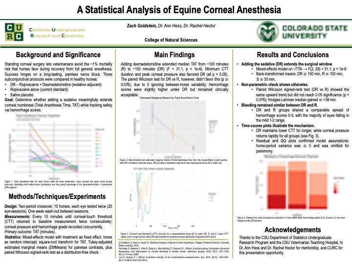
Effective local anesthesia of the equine cornea is essential for standing ophthalmic procedures and helps avoid the ~1% mortality risk of general anesthesia in horses. Ropivacaine is a long-acting local anesthetic that produces regional sensory loss. Dexmedetomidine, an α₂-adrenergic agonist with sedative and analgesic properties, was evaluated as an adjunct to determine whether it prolongs ropivacaine’s effect. In a randomized two-period crossover study, ten healthy adult horses (one eye excluded for cataract) received dexmedetomidine plus ropivacaine (DR), ropivacaine (R), or saline (S) in each eye under light sedation, with treatments switched after a one-week washout. Corneal touch threshold (CTT) was measured using a Cochet–Bonnet esthesiometer, and corneal pressure was measured by tonometry at baseline and every 10 minutes until two consecutive baseline CTT readings defined recovery. Total time under anesthesia (TTA) was defined as the interval from injection to return to baseline sensitivity. Injection-site hemorrhage was scored on a 0–4 scale. Linear mixed-effects models with horse as a random intercept were fit to √TTA, duration of minimum CTT, and maximum corneal pressure. Treatment significantly affected √TTA (F(2,29)=31.09, p=5.9×10⁻⁸), with DR>R (Δ=3.42; p=0.028), DR>S (Δ=8.43; p=6.9×10⁻⁸), and R>S (Δ=5.01; p=0.0002). DR prolonged maximal numbness and increased maximum corneal pressure. Hemorrhage scores were higher with active drugs than saline but did not differ between DR and R. These findings support dexmedetomidine as an adjunct to ropivacaine for prolonged corneal anesthesia.